Thematic focus and development of the Centre
The research activity of
the Centre has an interdisciplinary approach and concerns the comprehensive
characterisation of matter, particular the attention is put on detecting the heavy
elements contents and the role and estimation of elements ionicity, which is
crucial for estimation of the level of hazards. Over the past century the
numerous techniques have been developed which investigate, on an atomic scale,
the structure of the matter and which enable the physicists to describe with
increasing precision the fundamental interactions. The techniques based on
radiation and particles interaction with matter occupy a special place in these
investigations. Beams of photon, electron, proton, light ion or atom and
neutrons are of a special interest of the Centre.
A beam of well-defined and well-known energy and direction
propagates through the matter under study and interacts with its basic
constituents. Using several “radiation-probes”, different and complementary
information can be gain.
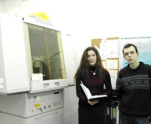
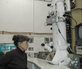
Photo
1.
High-resolution diffractometer Philips-MRD and PhD students.
q
HASYLAB at DESY, Hamburg, Germany,
q
L.U.R.E. , Universite de Paris-Sud,
Orsay, France,
q
Advanced Light Source, Lawrence
Berkeley Laboratory, Berkeley, USA,
q MAX-Lab, Lund University, Lund, Sweden,
q
ESRF, Grenoble, France and
q
ELETTRA, Trieste.
X-ray
diffraction and imaging methods help to diagnose several diseases like
osteoporosis and another bones illnesses. The investigation of crystal formed
in human body (e.g. bile or kidney stones) help with recognising the morphology
of the diseases. The Centre actively cooperates with medical clinics and
participates in the programs aimed to estimate the correlations between
pollutions of environment and content of heavy elements in bones, stones and
tissues. As an example, there was proposed the new complete method for analysis
of the microstructure of a trabecular bone in human bone bioptats. This method
is based on the special X-ray high resolution imaging the structure and the
mathematical method of its analysis by using the Fourier Transform of series
obtained X-ray images of bone, the fractal dimension analysis and finally the
discrimination analysis method. The correctness of the diagnoses was on the
level of 98%. The method has been realised in collaboration with the Institute
of Applied Optics - Warsaw, the Food and Nutrition Institute - Warsaw and the
Ortopedic Hospital - Otwock. Two doctors theses were prepared on the base of
this method.
For studies of chemical compositions and chemical states of the
elements the other advanced technique has been also applying in the Centre for
many years. This is the ESCA (electron spectroscopy for chemical analysis).
Starting from the post doctoral
position of the senior of the Centre prof. J. Auleytner at the Siegban
Laboratory in Uppsala, the several scientists
still continue the collaboration
with the Sweden and Finnish Laboratories equipped with the modern ESCA and
Auger spectrometers. The photoelectron spectroscopy is used for
elemental composition, chemical state, and band structure studies of crystal
surface and this method, using synchrotron radiation, is generally acknowledged
as the most useful tool for the band structure studies of crystals. Moreover,
it is widely applied in the Centre for investigations of the phenomena
occurring on the surface and in subsurface layers of solids. Together with
another techniques, it is successfully used at realisation of several research
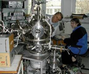
projects of the Centre. The self made UHV set-up assembled in the Institute of
Physics (Photo 6) enables us to carry out surface studies of solid samples. The
clean surfaces of the samples, suitable for surface studies, can be prepared by
cleavage under UHV conditions or by annealing in situ. The properties of
clean surfaces or of those modified by deposition of small amount of metals can
be investigated by means of low energy electron diffraction (LEED) and Auger
electron spectroscopy. The set-up is still developed in order to increase the
number of preparation and experimental techniques being at our disposal. In
particular, assembling of a system for surface sensitive optical measurements
is planed.
Finally, the
neutron particles are also applied in the Centre to sample the matter owing to
their very specific and unique properties. Neutron has no charge and has a spin
of ½ which is the equivalent to same value of a magnetic moment and,
consequently, is sensitive to the magnetic fields created by unpaired electrons
present in the material under studying. In magnetic materials the
matter-neutron interaction allows to study the magnetic order (magnetic
structure, magnetic phase transitions) and the possible excitations in magnetic
system, in technological materials as well in natural and biological samples. Selected neutron scattering techniques
enable to detect even small contamination of the matter investigating the chosen magnetic or heavy
elements, which is very important from the point of view of the environment
pollution studies. In cooperation with the Laboratoire Le’on Brillouin
(LLB) in the CE Saclay research center
the scientists from the Centre carry out
research programs covering different topics in the fields of condensed
matter physics, magnetism and superconductivity, physical chemistry, biology
and medicine as well as in material sciences (in particular, in the physical
metallurgy).
The Centre engages in its
activity more than 1/10 part of the Institute of Physics. It employs 43 staff
members among whom 36 are directly involved in the research activity. The
personal roster is the following: 2 full professors, 5 associate professors, 19
researchers with the doctor’s degree, 4 assistance, 6 Ph.D. students (2 from
Ukraine), and 7 technical staff.
The several former employees took the position of
the permanent staff abroad. They are employed at the scientific institutions in
the Federal Republic of Germany, the USA , Canada, Brazil, and Australia.
In summary, the
research activities of the Centre concentrate on the developing of modern
methods of characterisation of matter. These methods are applied with success
to the variety of physical problems in technology of modern materials. The
application to another natural materials (e.g. archaeological, geological,
medical biological and environmental) has already started in the Centre. We do
hope that with the help of UE grant it will be developed more intensively.
IN CONCLUSION,
the research activities in the Centre attempt to bring together all important
aspects of the modern methods of samples characterisation:
q
conductive and non-conductive
samples preparation and characterisation,
q
experimental studies of interaction
of beams with matter,
q
theoretical investigation,
including the basic aspects of interacting of beams with
matter,
q
computer simulation of the
interaction and comparison with the observation,
q
estimation of the confidential limits
of the evaluated characteristic parameters.